Sea Urchin Spine Removal
The Heat-First Rule. Why squeezing fragments the spine deeper, what hot water actually does to echinoderm venom, and the field protocol for removal without retained foreign body.

Your fingers are making it worse. The reflex when a sea urchin spine breaks off in the foot is to squeeze it out, to pinch the tissue around the entry point and try to drive the fragment back the way it came. That reflex is the single most common reason a clean field extraction turns into a months-long granuloma. Echinoderm spines are calcium carbonate. They are brittle, structured for puncture, and not structured to survive compression. The force from a thumb and forefinger does not lift the spine out of the dermis. It shatters the spine inside the dermis, leaving fragments distributed along the wound tract that the body cannot easily mobilize and that imaging may or may not find later.
This piece walks through what actually works at the break, in order, with the evidence each step rests on. The setting assumed throughout is the one most surfers and lifeguards meet this injury in: a remote reef, no immediate access to a clinic, time and tools limited to what is in the kit.
Why this injury behaves the way it does
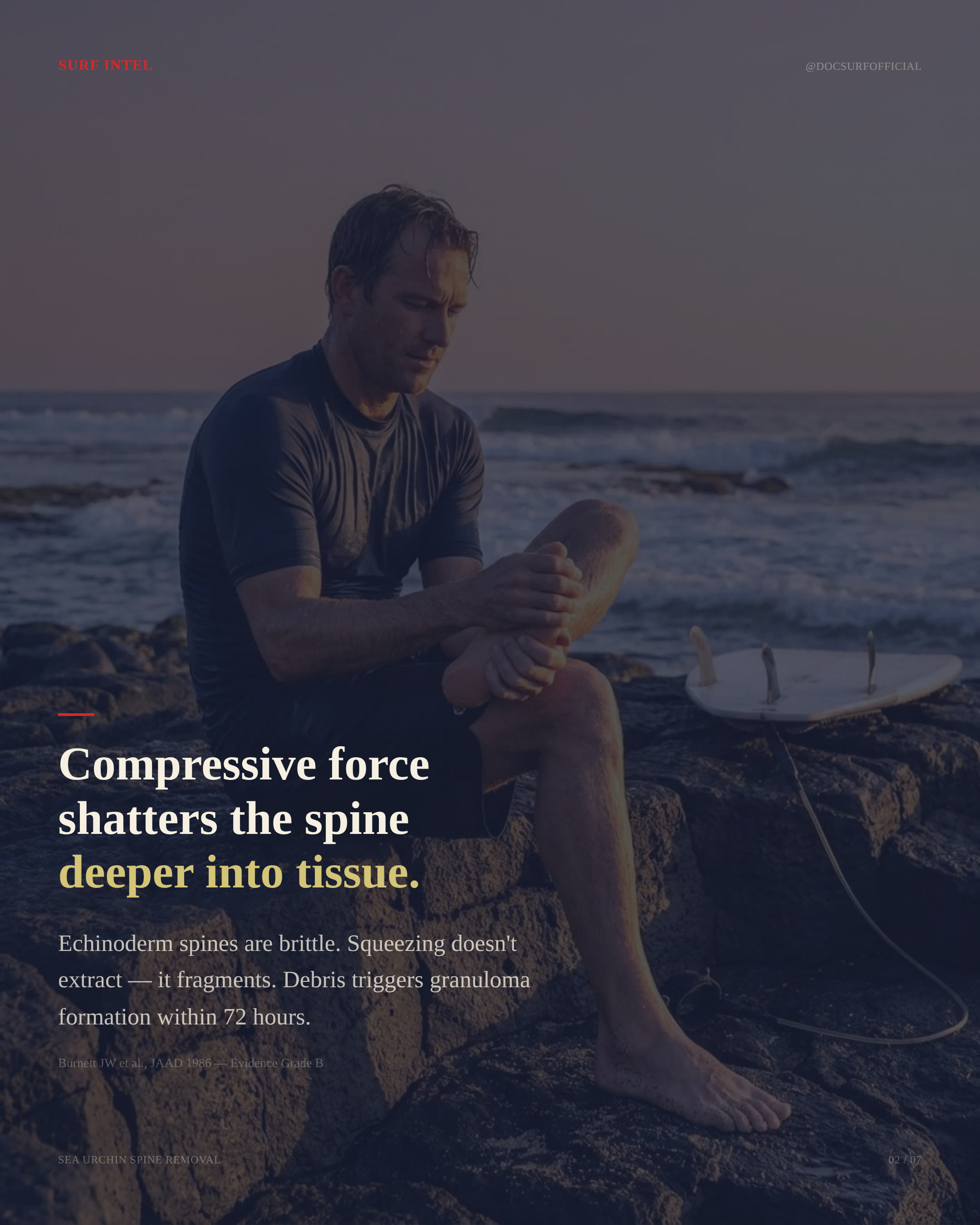
Sea urchin spines combine two problems in one puncture. The first is mechanical. The spine itself is a mineralized rod, often barbed along its length, designed by the animal for defense against soft-tissue predators. When a spine enters human tissue it deposits along the entry tract and tends to fragment under any force that is not aligned with that tract. The second problem is chemical. Many reef-dwelling urchin species coat their spines in venom proteins delivered through pedicellariae or through the spine surface itself. These proteins drive the disproportionate pain that follows what looks, on first inspection, like a small puncture.
Both problems shape the response. The mechanical problem means extraction technique is not optional finesse, it is the difference between a clean removal and a retained foreign body. Fragments left in tissue cause foreign body granuloma within 72 hours, a low-grade inflammatory reaction that organizes around the retained material and can persist for weeks to months before the body either walls it off or pushes it out (Burnett et al., 1986). The chemical problem means that pain control and tissue softening can be addressed before the extraction is attempted, which makes the extraction itself easier and cleaner.
The protocol, in order
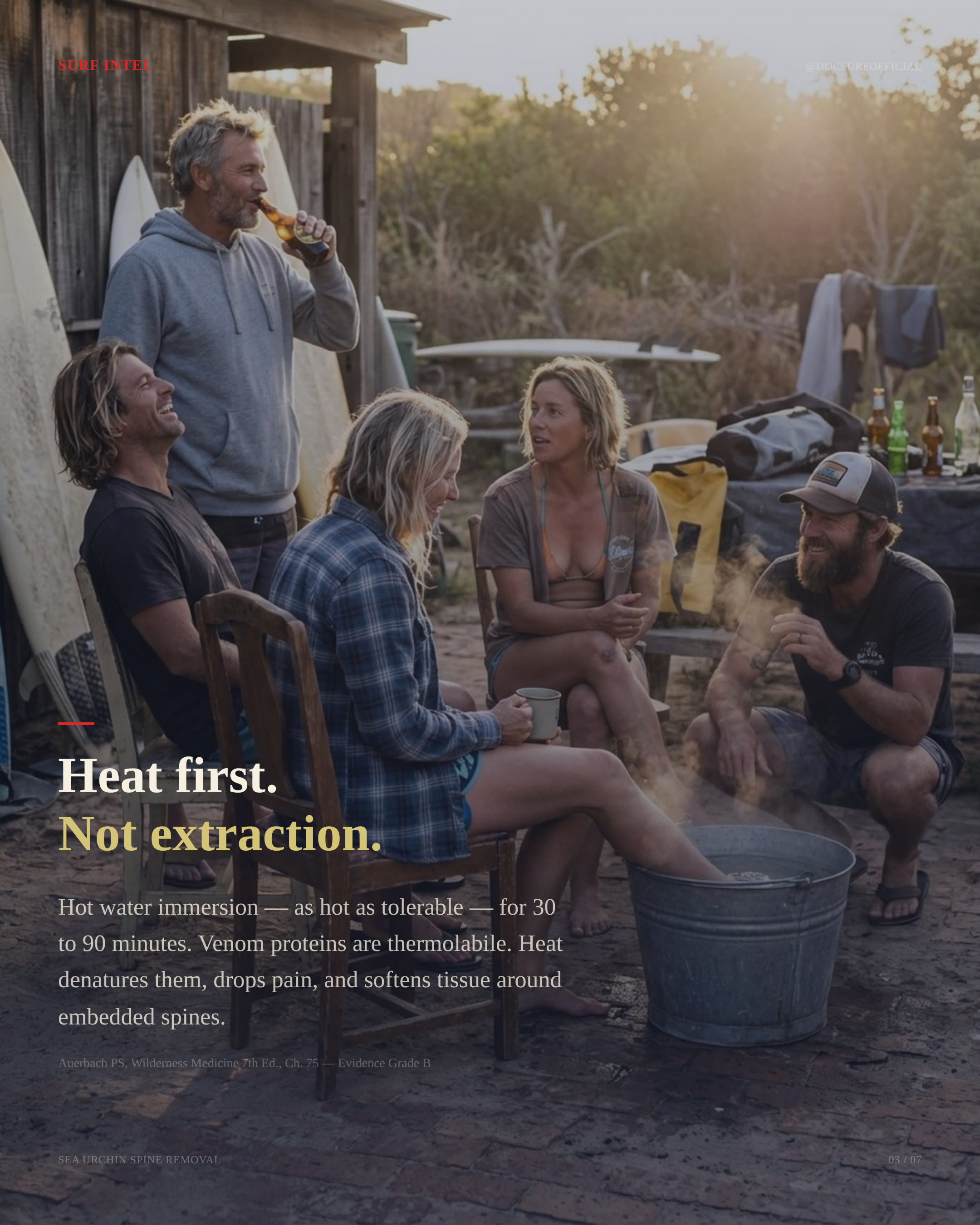
Step one is heat, not extraction. Before the tweezers come out, the affected area goes into hot water. The target range is 43–45°C, as hot as the patient can tolerate without scalding. Immersion time is 30 to 90 minutes. The mechanism is straightforward: the venom proteins driving the pain are thermolabile, meaning heat denatures them. Pain drops significantly inside the first ten to twenty minutes for most patients. A secondary effect of the immersion is that the tissue surrounding the embedded spine softens, which reduces the resistance against the spine during extraction and lowers the force required to lift it out cleanly (Auerbach, 2017, Ch. 75).
The temperature ceiling matters. Water hotter than 45°C risks a thermal burn layered on top of the puncture, which compounds the injury and complicates wound management. A reliable field method is to test the water with the uninjured limb of the patient or with a kit thermometer before the affected area goes in. If neither is available, the rule is that the water should be hot enough to be uncomfortable but not hot enough that the patient pulls away within seconds.
Step two is fine-tip tweezers, aligned parallel to the angle of entry. Not fingernails. Not a sewing needle pushed in alongside the spine. Not a credit card scraped across the surface. The geometry is the entire point: the spine entered along a specific vector, and the only force that lifts the spine out without fragmenting it is force applied along that same vector. Oblique force, lateral force, or compressive force fractures the spine along its length and seeds fragments deeper into the wound tract (Perrine, 2016; Borden Institute, 2013).
Fine-tip tweezers give the operator the precision to grip the spine at its exposed end without crushing the shaft. Wider tweezers, or any improvised instrument that grips the spine broadly, function essentially the same way as a thumb and forefinger and produce the same fragmentation pattern. If fine-tip tweezers are not in the kit, the protocol calls for delaying extraction until they are available rather than attempting it with the wrong instrument.
For calcium carbonate spines that do not come out cleanly after heat immersion and parallel extraction, topical marine sting neutralizer applied to the wound dissolves the mineral structure of the spine over time (Auerbach, 2017, Ch. 75). This is a slower process measured in hours, not minutes, and it is not a substitute for the initial extraction effort. It is the option for fragments that the tweezers could not retrieve, or for spines so deeply embedded that the field operator judges further extraction attempts more likely to cause tissue damage than to recover the fragment.
Leave the wound open. Marine puncture wounds, particularly those acquired in warm reef water, carry a high background risk of Vibrio vulnificus contamination. V. vulnificus is ambient in warm reef water, in sediment, and in the surface mucus of coral itself (Diaz, 2005). Closing the wound with adhesive strips, tissue glue, or sutures in the field traps the organism in an anaerobic environment with damaged tissue to feed on, which is the precise condition that V. vulnificus exploits. The wound is irrigated, packed open, dressed loosely, and monitored.
Where the protocol goes wrong
The field-common errors cluster in three places.
The first is skipping heat. The instinct under stress is to go straight to extraction, because the spine is visible and the patient is in pain and the operator wants to act. Extraction without prior heat immersion is harder, more painful, and more likely to fragment the spine, because the tissue is still tight around the foreign body and the venom proteins are still active. The thirty to ninety minute immersion window feels long when the patient is uncomfortable and the group wants to keep moving, but it is the step that makes every step after it work.
The second is using the wrong tool. Fingernails, sewing needles, knife tips, and improvised grippers all produce the same outcome: fragmentation. The correct tool is fine-tip tweezers, and the correct kit contains them. A reef-going first aid kit without fine-tip tweezers is a kit that cannot manage the single most common reef puncture cleanly.
The third is closing the wound. Surfers and lifeguards trained primarily in clean-environment first aid sometimes treat a marine puncture the way they would treat a clean laceration from a kitchen knife. The marine environment is not clean. V. vulnificus risk is the dominant consideration in any reef puncture, and wound closure in the field is contraindicated under that risk profile.
When this leaves the field
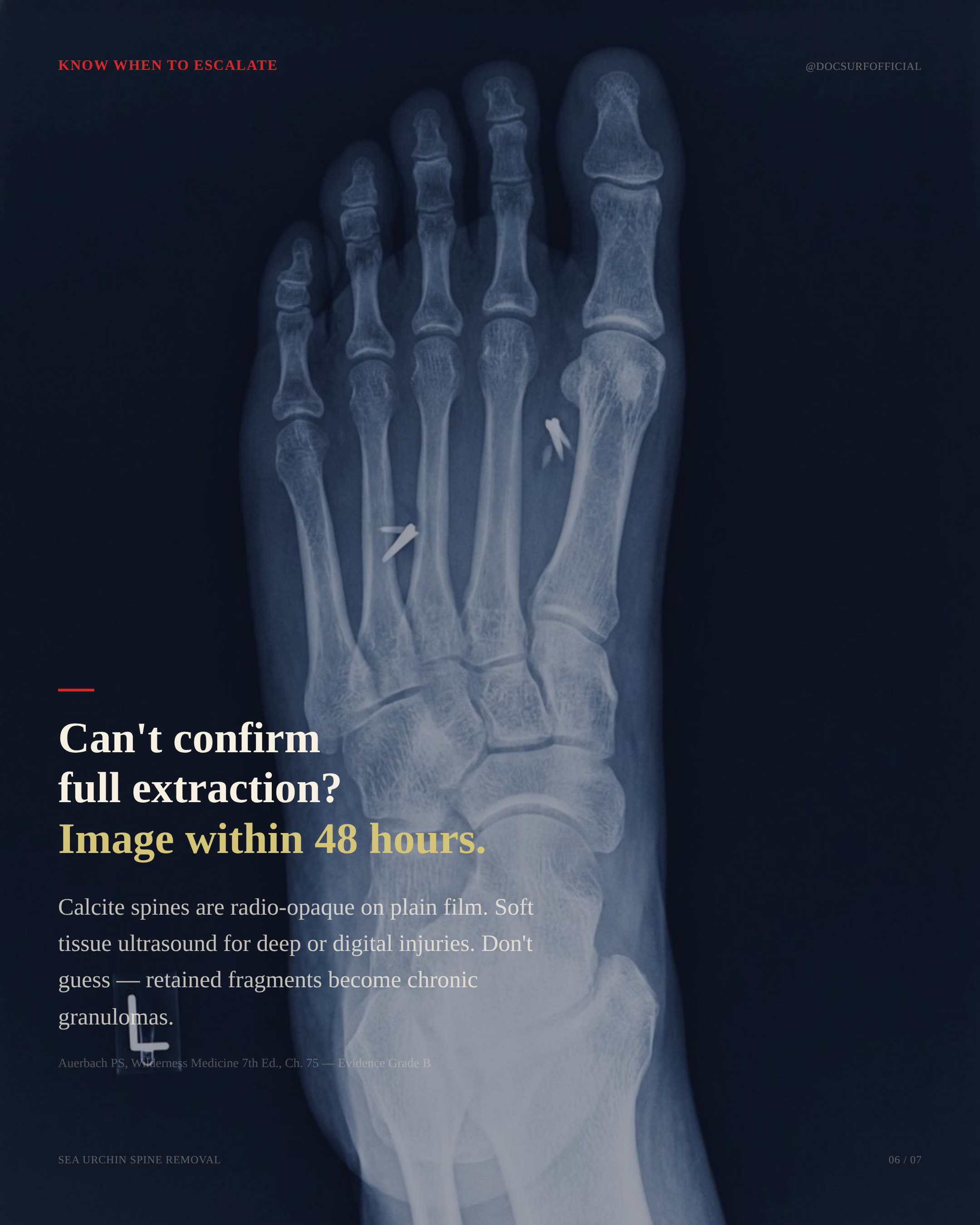
If complete extraction cannot be confirmed in the field, the next step is imaging within 24 to 48 hours. Calcite spines are radio-opaque on plain film, which means a standard x-ray will typically locate retained fragments in the foot, hand, or other extremity. For deep or digital injuries, where the bony anatomy can obscure small fragments on plain film, soft tissue ultrasound is the more sensitive option and is increasingly accessible in regional centers.
The threshold for imaging is conservative. A patient who clearly extracted the spine intact, with the full spine recovered and accounted for, can reasonably defer imaging and watch the wound. A patient with any uncertainty about whether the full spine was recovered, or any sign of fragmentation during the extraction, falls into the imaging window. Delayed presentation of retained fragments as chronic granuloma, persistent pain, or recurrent local infection is common enough that the field protocol errs toward earlier imaging when there is doubt.
Escalation also applies to the wound itself. Marine puncture wounds that show expanding erythema, disproportionate pain after the first 24 hours, fever, or any sign of systemic involvement leave the field protocol and require evaluation by a qualified provider. V. vulnificus infection moves fast in vulnerable hosts, and the window between local infection and systemic involvement can be measured in hours rather than days.
What to carry
The kit for this injury is small. Fine-tip tweezers. A reliable means of heating water to 43–45°C and holding it there for up to ninety minutes, which in most field contexts means a stove and a vessel large enough to immerse a foot or a hand. A thermometer that reads into that range. Topical marine sting neutralizer for fragments that resist initial extraction. Irrigation supplies and loose dressings for the open wound. A way to document the time of injury, the time of immersion, and what was recovered, because that information matters at the imaging stage if the wound goes that way.
The protocol itself is short enough to carry in the head. Heat first, for 30 to 90 minutes at 43–45°C. Fine-tip tweezers, parallel to the angle of entry. Neutralizer for stubborn fragments. Leave it open. Image within 24 to 48 hours if extraction is uncertain. The errors that turn a clean reef puncture into a chronic foreign body problem are almost all errors of sequence and tool selection, not errors of knowledge. The medicine is not complicated. The discipline is in doing the steps in the right order, with the right instrument, under conditions that push the operator to skip both.
References
- Burnett JW, Calton GJ, Burnett HW. Local and systemic reactions from jellyfish stings and sea urchin envenomations. Journal of the American Academy of Dermatology. 1986.
- Auerbach PS, ed. Wilderness Medicine. 7th ed. Philadelphia: Elsevier; 2017. Chapter 75: Envenomation by Aquatic Invertebrates.
- Perrine D. Sea Urchin Envenomation and Injury: Clinical Review and Field Management. 2016.
- Borden Institute. Textbook of Military Medicine: Medical Aspects of Harsh Environments. Washington, DC: Office of the Surgeon General, United States Army; 2013.
- Diaz JH. The epidemiology, diagnosis, management, and prevention of marine wound infections. Journal of the Louisiana State Medical Society. 2005.